 Pellaeon
A ligand in its pocket
HIV-1 protease with the inhibitor indinavir (PDB 1HSG), in four requests.
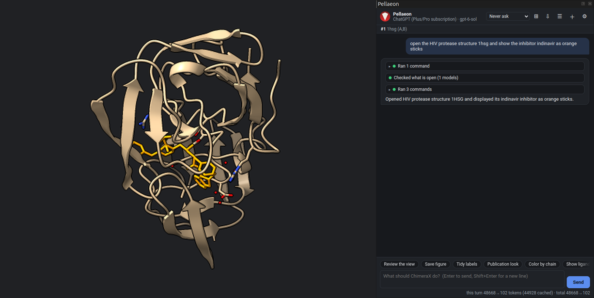
1. Open the structure
open the HIV protease structure 1hsg and show the inhibitor indinavir as orange sticks
open 1hsg
show #1:MK1 atoms; style #1:MK1 stick; color #1:MK1 orange
MK1 is the residue name of indinavir in this entry.
2. The pocket as a surface
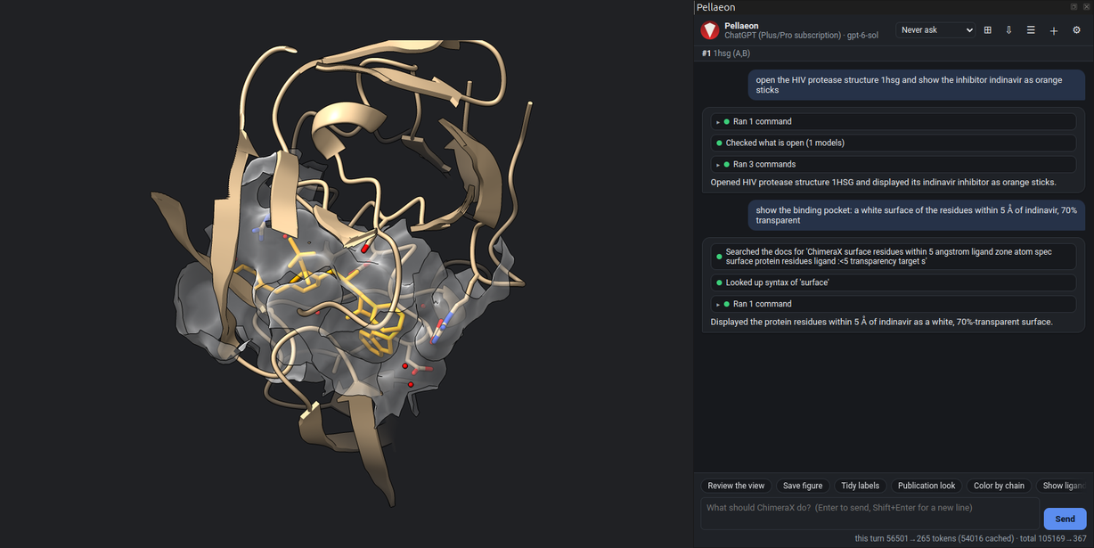
show the binding pocket: a white surface of the residues within 5 Å of indinavir, 70% transparent
surface (#1:MK1 :<5) & protein color white transparency 70
:<5 selects the residues with any atom within 5 Å of the ligand.
3. Hydrogen bonds
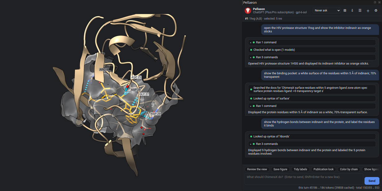
show the hydrogen bonds between indinavir and the protein, and label the residues it binds
hbonds #1:MK1 restrict protein reveal true select true
select sel & protein; label sel
Nine hydrogen bonds to five residues: Arg 8, Asp 25 of both chains, Gly 27 and Asp 29. The catalytic aspartates (Asp 25) bind the inhibitor's central hydroxyl.
4. A close-up
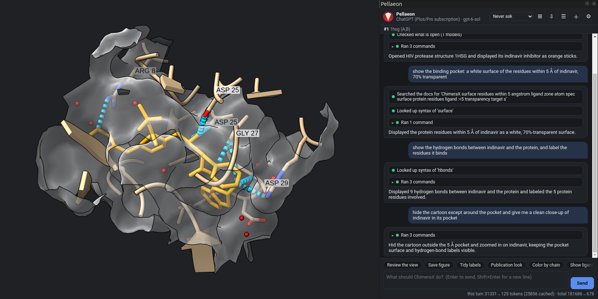
hide the cartoon except around the pocket and give me a clean close-up of indinavir in its pocket
hide #1 target c
cartoon (#1:MK1 :<5) & protein
view #1:MK1
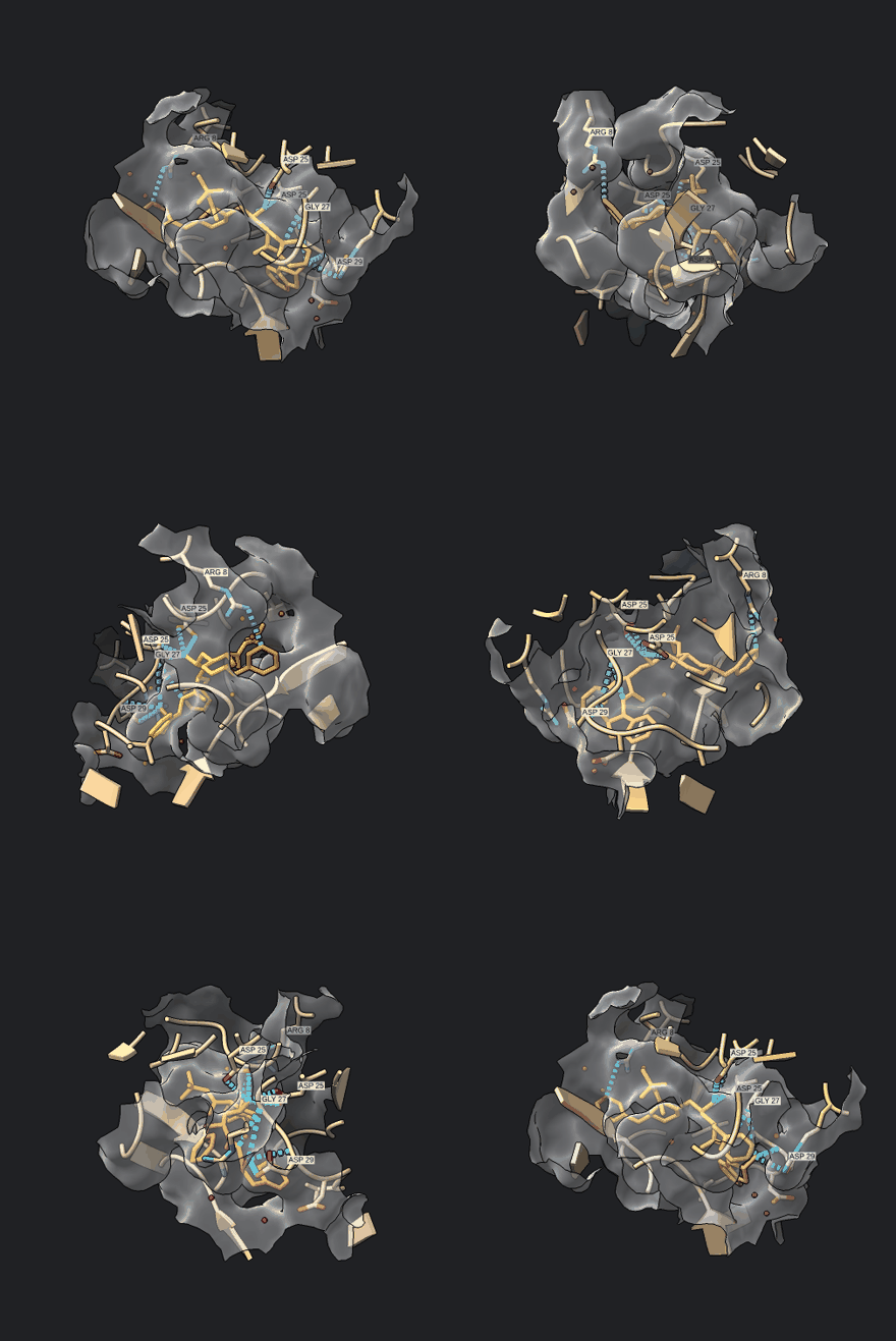
Save figure writes the image, the session and the commands to a folder (tutorial, step 5b).