 Pellaeon
Electrostatics and hydrophobicity
The p53 core domain bound to DNA (PDB 1TSR), in four requests.
1. Open the structure
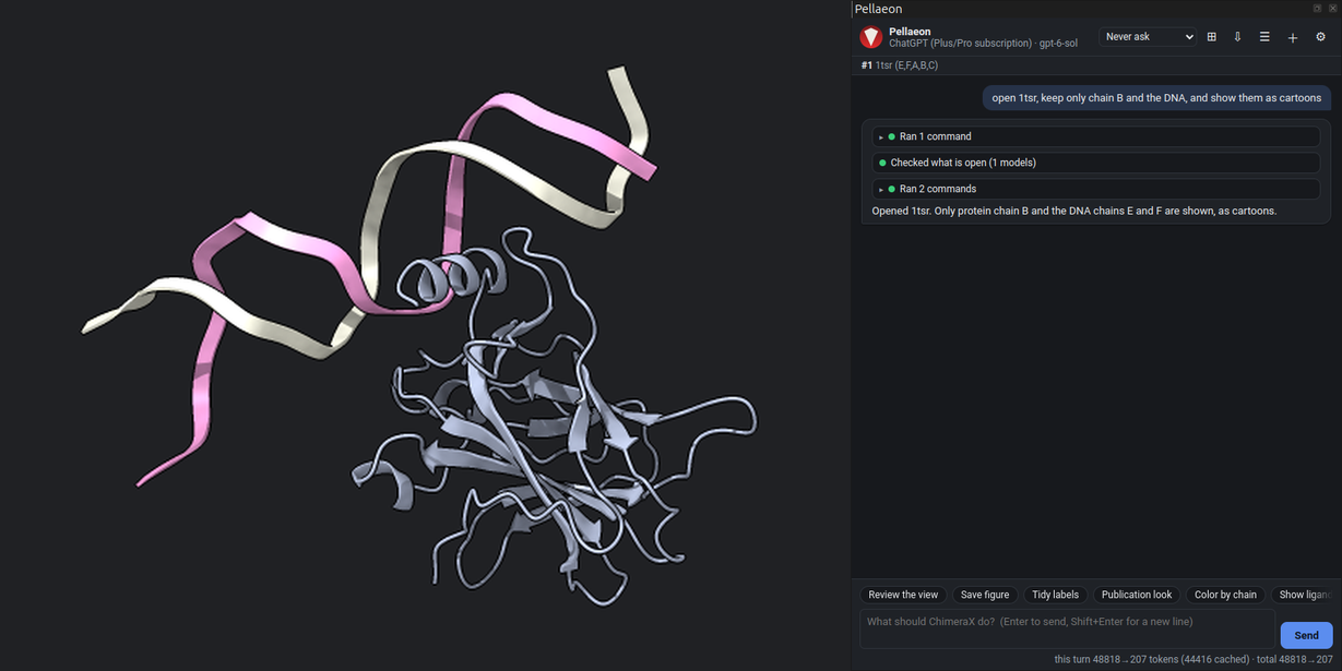
open 1tsr, keep only chain B and the DNA, and show them as cartoons
open 1tsr
hide #1 target acs; cartoon #1/B,E,F
The entry has three copies of the domain (chains A, B, C); chain B is the one on the DNA (chains E and F).
2. Electrostatic potential
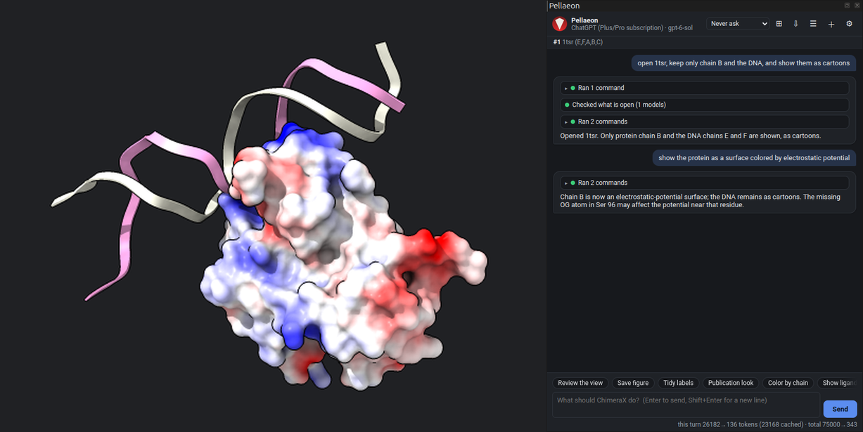
show the protein as a surface colored by electrostatic potential
cartoon hide #1/B
coulombic #1/B
Red is negative, white neutral, blue positive. ChimeraX warns that Ser 96 lacks an atom, so the potential near it is approximate.
3. Hydrophobicity
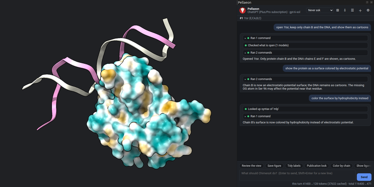
color the surface by hydrophobicity instead
mlp #1/B surfaces #1.3
Molecular lipophilic potential on the same surface: hydrophobic patches orange, polar ones teal.
4. Which face binds the DNA
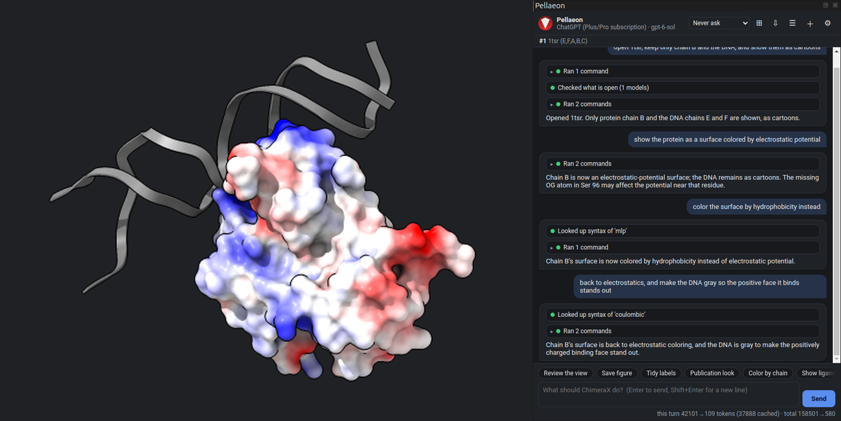
back to electrostatics, and make the DNA gray so the positive face it binds stands out
coulombic #1/B surfaces #1.3
color #1/E,F gray
The positively charged face of the domain lies against the phosphate backbone.